


Thoriax – PC ECG
General description
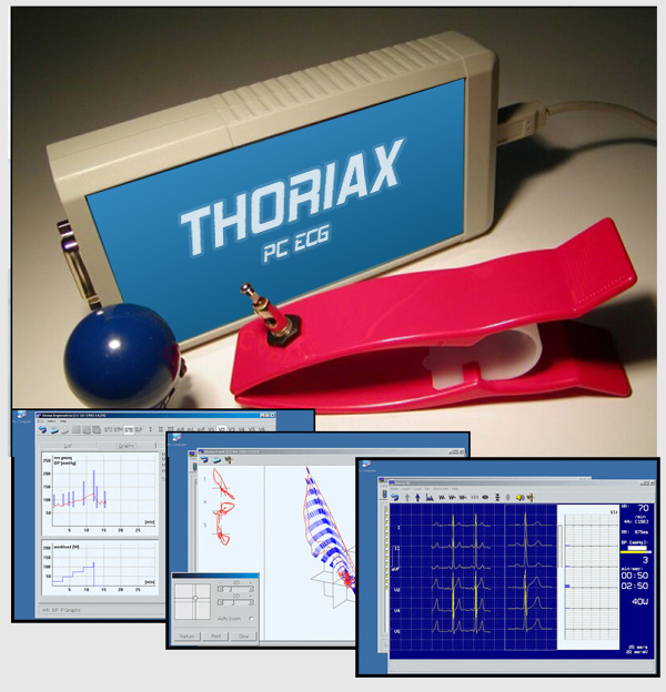
Thoriax is a PC-based computerized electrocardiograph system with full ergometry functionality.
Thoriax consists of three main parts:
- The ECG sampler is a very small external computer peripheral device that can be connected to any IBM PC-based computer via USB or parallel port.
- The electrodes are all connected to the ECG sampler device.
- Visualization and recording are performed by the Thoriax software that is a 32-bit application running on all versions of Microsoft Windows.
Thoriax is a high quality 12-lead ECG system. Thoriax is extendable with a number of medical systems (ergometers, blood pressure meters, suction electrodes, etc).
Specification
| Sampling | 2000 samples/sec |
| Resolution | 12 bits |
| Number of channels | 12 |
| Input impedance | > 10 Mohms |
| Patient protection | floating earth (IEC 601) |
| Defibrillator protection | 5 kV (1 kV/microsec) |
| CMRR | >120 dB |
| Filter | 35, 50/60, 100/120 Hz |
| Time constant | 3.2 sec |
| Linearity error | <0.5 %, < 1/2 LSB |
| Polarization voltage | ą400 mV |
| Leads | 12 standard + 3 Frank |
THORIAX stress test
The THORIAX stress test module fully automates stress tests. The computer manages full control of the stress equipment, a bicycle or a treadmill. The whole process of stress testing is pre-programmed. You can preset phase duration, initial load and load steps, as well as the conditions to break stressing. If the ergometer is equipped with an automatic blood pressure meter, the THORIAX system will read it and store the measured values. An ECG segment is automatically saved at the end of each stress phase. Interpretation is performed on the fly, so median curves and ST-trends can be shown on the display while the test is in progress.
When the stress test is finished, a final report is composed of the results. You may review the results of the test in graphical format too. The final graphs contain the heart rate, blood pressure and load by time. Other graphs show the ST amplitudes and medians by time (ST trend and QRS trend). The program also evaluates the physical work capacity in the maximal and submaximal range.
THORIAX filer
The personal data, ECG recordings, diagnoses and other data of patients are stored in the THORIAX filer. Finding someone in the list of patients is quick and easy. The list can be sorted by name, patient code, age of patients and the date of the last recording. You can select patients in the filer meeting some conditions simply filling out a simple form. The card and recordings of selected patients can be archived to floppies or CD-ROM, or copied to and from other filers. The THORIAX program is an open system. It can easily be connected to third party medical databases and patient software.
The THORIAX system can be installed on a LAN too. In this case one workstation can serve as an ECG recording equipment, while other workstations can be used to retrieve and examine existing ECG recordings.
THORIAX as an ECG monitor
While monitoring, the running ECG curves are shown on the computer display. Changing the number of channels (3,6,12), paper velocity (up to 400 mm/s), the amplification and the lead combination is just a keystroke away. Beside the standard lead systems (e.g. Standard, Frank, Precordial, Cabrera etc.) you can freely define any 3 or 6 channel lead combination. You can store segments of 8, 16 or 32 seconds at any time.
THORIAX printing
Patient information, ECG strips, median curves and diagnoses can be printed on any IBM PC compatible standard printer.
THORIAX interpretation
The interpretation module uses data of the 12 channels to calculate the averaged QRS complexes (medians) and the rhythm strip. This module locates the diagnostically important parameters. Based on its analysis, the program offers a suggested diagnosis for the doctor, and stores it in the filer. The system can diagnose adult patients (over 15), as well as children (2-15 years old).
THORIAX viewers
The stored ECG recordings can be viewed in any lead combination. It can be examined statically, by paging the recording to and fro, or can be replayed exactly the same way as it was recorded. The static curves can be used to make measurements with the help of markers.
You can select some recordings and compare them in a single ECG window. You can also compare the medians of multiple selected recordings. The selected recordings can be resting ECG recordings or recordings of a stress test.
For the recordings of Frank lead combination the ECG vector of a selected curve segment can be viewed as a vector cardiogram (VCG). The VCG is shown with it’s two dimensional projections and a three dimensional view. The 3D view can be zoomed, panned and rotated around in any direction.


